


Why does carrying APOE4 increase the risk of Alzheimer’s disease? Despite decades of research, we still do not have a complete answer.
APOE4 is the strongest common genetic risk factor for late-onset Alzheimer’s disease, but its effects extend beyond cholesterol transport. Research suggests that APOE4 alters how brain cells handle fats, affects blood vessel function, and makes neurons more vulnerable to injury; we have studied how APOE4 impairs the resolution of neuroinflammation by looking at omega-3 and omega-6 metabolism.
Our laboratory studies have also examined another aspect of APOE4 biology: cholesterol metabolism and the formation of lipidated particles. One important question is whether improving APOE4’s ability to transport cholesterol could reduce some of its harmful effects.
Our latest study, led by Zoe Tsokolas and published last week in Alzheimer’s Research & Therapy, examines one part of this larger question. We compared small and large HDL particle concentrations with APOE protein concentrations, measuring both in blood and cerebrospinal fluid, the fluid surrounding the brain and spinal cord, in an extremely well-characterized cohort. We then investigated how these measurements related to Alzheimer’s biomarkers. [1]
The central finding was that APOE4 protein concentration in cerebrospinal fluid was more strongly associated with tau pathology than the measured numbers of HDL particles.
To understand why this matters, we first need to explain how APOE normally transports fats in the brain.
What does it mean to lipidate APOE?
The brain needs cholesterol and other fats, collectively called lipids, to maintain cell membranes, repair damage, and support communication between neurons.
One of the main proteins that transports these fats between brain cells is apolipoprotein E (APOE), produced mainly by support cells called astrocytes.
APOE combines with cholesterol and other lipids to form particles called lipoproteins. The process of attaching lipids to APOE is known as lipidation.
These particles resemble high-density lipoproteins, or HDL, although brain HDL differs from the HDL commonly measured in blood cholesterol tests.
HDL particles vary in size and composition. Small HDL particles carry relatively little lipid and can exchange cholesterol with cells. As they acquire additional lipids, they can become larger HDL particles.
A protein called ABCA1 helps initiate lipidation by transferring cholesterol and other lipids from cells onto APOE. Other proteins can contribute to further lipid loading.
Why does this matter for Alzheimer’s disease?
In animal models, increasing functional ABCA1 activity can improve APOE lipidation and reduce amyloid accumulation in the brain. Amyloid is one of the abnormal proteins associated with Alzheimer’s disease. [2]
These findings led to an important hypothesis: improving APOE lipidation might help protect the brain.
How does APOE4 interfere with lipid transport?
There are three common forms of APOE: APOE2, APOE3, and APOE4. Each person inherits two copies of the APOE gene.
Our group previously demonstrated that APOE4 interferes with the normal movement of ABCA1 inside astrocytes. [3]
ABCA1 must reach the cell surface to help transfer cholesterol onto APOE. In 2019, we reported that with APOE4, more ABCA1 becomes trapped in compartments called late endosomes and lysosomes, which normally sort, recycle, and break down cellular materials [3].
Consequently, less ABCA1 reaches the cell surface, cholesterol transport becomes less efficient, and more APOE remains poorly lipidated.
Poorly lipidated APOE4 is also more likely to form abnormal protein aggregates, or clumps.
Experimental treatments that restored ABCA1 activity improved some of these abnormalities, supporting the possibility that correcting lipidation could be beneficial.
However, our 2025 study revealed an important complication. [4]
In Alzheimer’s brain tissue and experimental models, ABCA1 levels could actually be increased, even though cholesterol handling remained abnormal. Much of the protein was trapped inside lysosomes rather than functioning at the cell surface.
This abnormal accumulation was associated with cholesterol buildup, altered cellular signaling, and features of cellular senescence, a stress-related state that can contribute to persistent inflammatory activity.
Simply increasing ABCA1 abundance may not improve cholesterol transport. The protein must also reach the right location and function properly.
These studies support continued investigation of APOE lipidation while reminding us that APOE4 affects several biological processes.
What question did our new study address?
Our earlier research found that older adults with higher concentrations of small HDL particles in cerebrospinal fluid (CSF) tended to perform better on cognitive tests. [2]
We called this the small HDL hypothesis of Alzheimer’s disease, and speculated that making more small HDL particles in the brain would be protective.
But there was a question we had not answered:
Were differences in Alzheimer’s biomarkers more closely related to HDL particle concentrations or to the amount of APOE protein?
To investigate this question, we analyzed samples from 144 participants in the Alzheimer’s Disease Neuroimaging Initiative (ADNI), a long-running research program studying cognitive aging and Alzheimer’s disease.
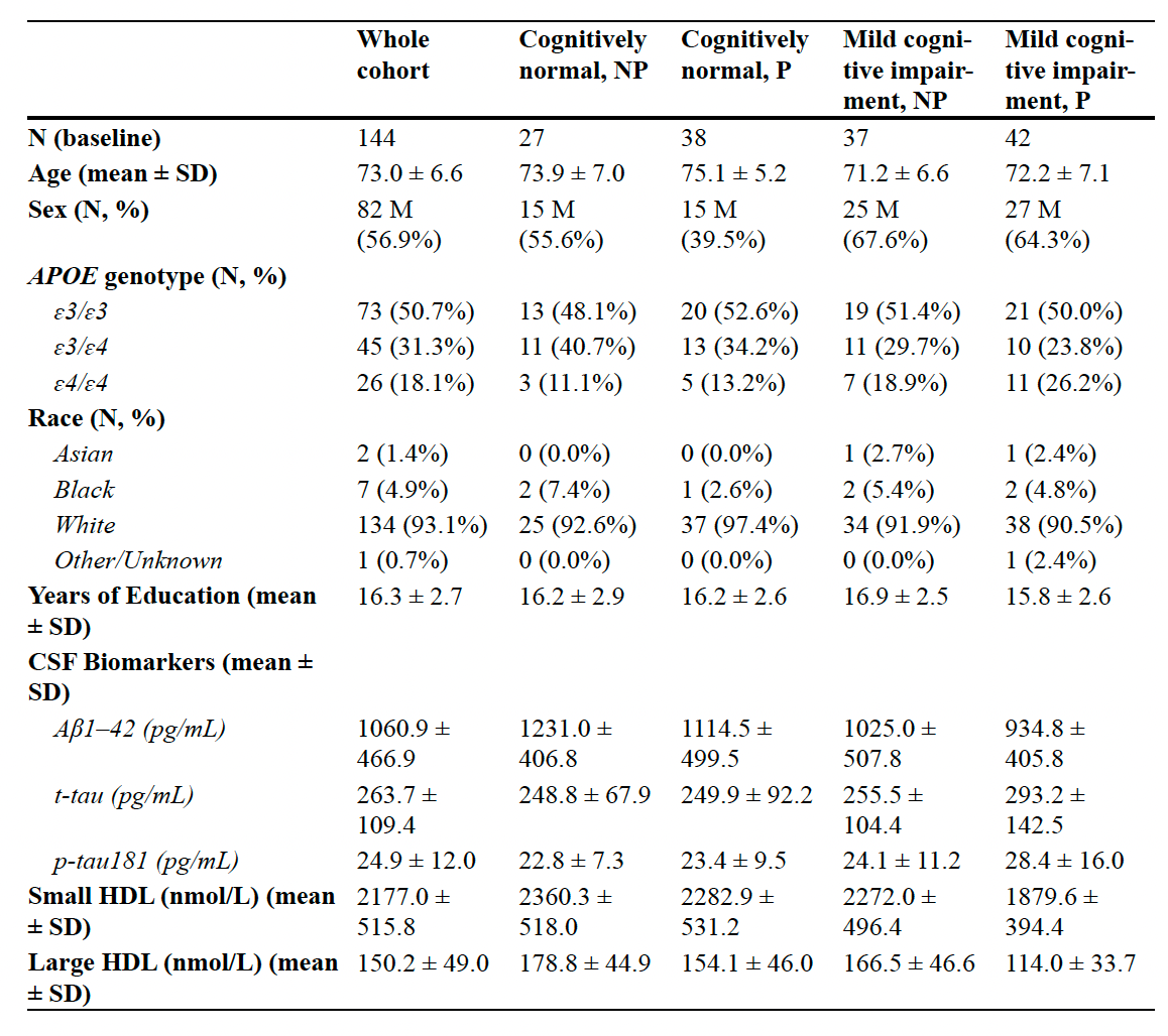
Participants were approximately 73 years old and included people with normal cognition and others with mild cognitive impairment (MCI)—measurable difficulties with memory or thinking that do not necessarily cause major loss of independence.
The group included 73 participants with APOE3/3, 45 with APOE3/4, and 26 with APOE4/4.
We also examined memory changes over four years.
What did we measure?
We used two complementary laboratory methods.
Ion mobility (IM) measured the concentrations of small and large HDL particles by separating them according to size.
Mass spectrometric immunoassay (MSIA) distinguished APOE3 from APOE4 protein and measured their relative abundance.
That distinction is particularly useful for people carrying APOE3/4, who produce both proteins.
Total APOE was measured in all 144 participants, while separate APOE3 and APOE4 measurements were available in 85 participants.
We compared these measurements with established CSF biomarkers, particularly total tau and phosphorylated tau (p-tau181).
Tau normally helps maintain the structure of neurons. In Alzheimer’s disease, tau undergoes abnormal changes and accumulates inside brain cells. Elevated CSF tau and p-tau181 are indicators of neuronal injury and Alzheimer’s-related pathology.
We wanted to know which measurements were most strongly associated with these markers.
What did we find?
1. APOE4 carriers had fewer small HDL particles in CSF
People carrying one or two copies of APOE4 had significantly lower concentrations of small HDL particles than those with APOE3/3.
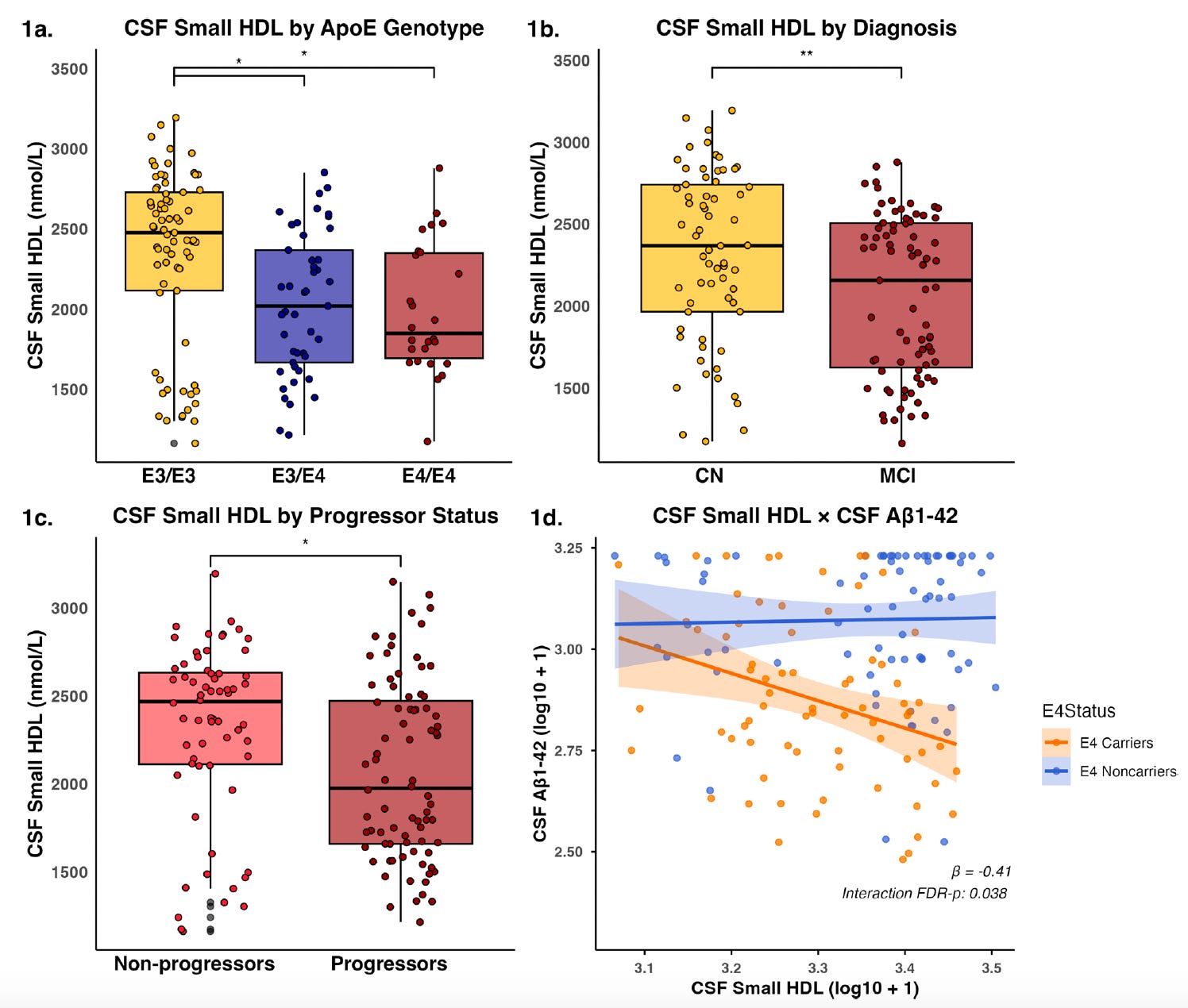
However, differences in large HDL concentrations across APOE genotypes were not statistically significant.
This is consistent with our earlier work showing that APOE4 disrupts normal lipid transport.
2. People with cognitive impairment had fewer small and large HDL particles
Participants with MCI had lower concentrations of both small and large HDL particles in CSF than cognitively normal participants.
The same pattern appeared when comparing participants whose memory or clinical status worsened over four years with those who remained more stable.
These observations suggest that changes in HDL particles accompany cognitive impairment.
But they cannot tell us whether reduced HDL contributes to disease or occurs as a consequence of it.
3. APOE4 protein concentration was more strongly associated with tau
This was the most important finding.
Higher CSF APOE4 protein concentrations were associated with higher concentrations of total tau and p-tau181. [1]
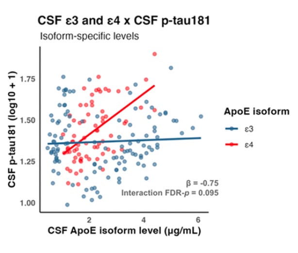
We then examined whether adding HDL particle measurements improved our ability to account for differences in tau.
In most analyses, they did not.
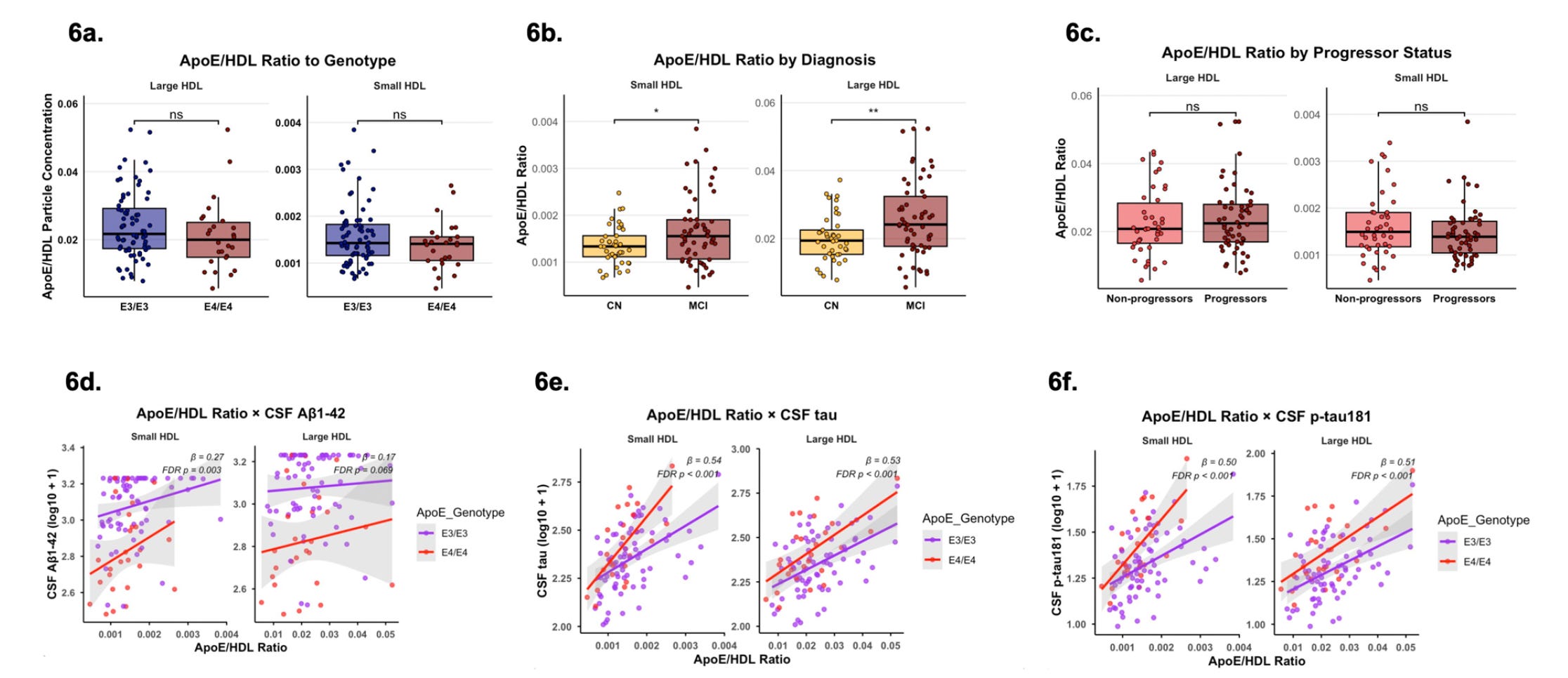
In the figure above, we examined whether the amount of APOE relative to the number of small or large HDL particles was associated with Alzheimer’s pathology. Participants with mild cognitive impairment had higher APOE-to-HDL ratios, and higher ratios were strongly associated with greater tau and phosphorylated tau levels in cerebrospinal fluid.
However, an important finding emerged when we compared statistical models: APOE protein concentration alone explained more of the differences in tau than the APOE-to-HDL ratio, and adding HDL particle numbers provided little additional information. This reinforces our central finding that APOE protein abundance may be more closely linked to tau pathology than HDL particle numbers. Importantly, the ratio does not directly measure how well APOE is lipidated or transports cholesterol. Therefore, these results do not diminish the potential value of improving APOE lipidation, but suggest that targeting the amount and harmful effects of APOE4 itself may be equally important, or perhaps more important, for addressing tau-related pathology.
Knowing the APOE concentration provided more information about tau pathology than knowing the number of small or large HDL particles.
This does not mean HDL particles are unimportant. Some HDL measurements were associated with Alzheimer’s biomarkers, and small HDL provided additional information about amyloid-beta.
Rather, the findings suggest that HDL particle abundance alone does not explain the relationship between APOE4 and tau pathology.
Why are people with APOE3/4 particularly informative?
People with APOE3/4 inherit one APOE3 gene and one APOE4 gene. They therefore produce both protein forms.
But the amounts are not necessarily equal.
In our study, APOE4 protein was more abundant than APOE3 protein in CSF among these participants.
We also found that the APOE4-to-APOE3 ratio was higher in participants with MCI than in those with normal cognition. [1]
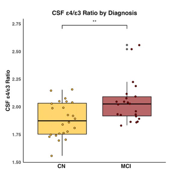
This raises an interesting possibility: people with the same APOE genotype may have different amounts of APOE4 protein, potentially influencing their biological vulnerability.
However, the APOE4-to-APOE3 ratio was not significantly associated with tau within the APOE3/4 subgroup.
The finding therefore raises a question rather than providing a definitive answer:
Could measuring APOE4 protein reveal information about Alzheimer’s biology that genetic testing alone cannot provide?
What did we learn from the bloodstream?
We also examined APOE and HDL particles in plasma, the liquid component of blood.
This matters because blood APOE and brain APOE are largely produced in different places.
Most circulating APOE comes from outside the brain, particularly the liver, while brain APOE is produced mainly by local cells.
In our study, total plasma APOE was lower in APOE4 carriers than in participants with APOE3/3.
However, total plasma APOE did not differ significantly between participants with normal cognition and those with MCI.
We measured small and large HDL particles in plasma as well, but the study did not establish the same pattern of significant HDL reductions seen in CSF.
Most importantly, plasma APOE measurements were not strongly or consistently associated with the Alzheimer’s biomarkers measured in CSF. [1]
This suggests that blood APOE measurements do not necessarily reflect what is happening in the brain.
Why might low plasma APOE still matter?
Large population studies have found that people with lower circulating APOE concentrations have a higher risk of dementia. [5,6]
These findings suggest that APOE in blood may have important protective functions.
However, they do not establish that increasing blood APOE prevents Alzheimer’s disease.
There is also no contradiction between these observations and our new findings.
Improving APOE function in blood and reducing harmful APOE4 in the brain are different biological strategies.
Both may deserve investigation.
Could improving APOE lipidation protect the brain?
Our group continues to investigate treatments designed to improve APOE lipidation.
One is CS6253, an experimental peptide developed by Artery Therapeutics that enhances ABCA1 activity.
In APOE4 mouse models, CS6253 improved APOE lipidation and reduced several disease-related abnormalities. [3]
In a subsequent study in monkeys, CS6253 increased small HDL particles and APOE concentrations in plasma and produced temporary changes in circulating amyloid-beta measurements. [7]
One possibility is that improving lipidation helps stabilize circulating APOE, allowing it to participate more effectively in lipid transport.
Another possibility involves the peripheral sink hypothesis.
This theory proposes that changing how amyloid-beta is handled in blood might favor its removal from the brain.
However, this remains unproven for CS6253.
In the monkey study, CS6253 produced clear changes in plasma but did not significantly alter the measured CSF lipoproteins. Very little peptide was detected in CSF relative to plasma.
Changes in blood amyloid measurements also do not establish that amyloid has been removed from the brain.
Improving peripheral APOE function is a plausible approach, but whether it protects the brain remains uncertain.
Should we also try to lower APOE4 in the brain?
Our new findings strengthen the rationale for investigating an additional strategy: directly reducing harmful APOE4 in the central nervous system.
If higher CSF APOE4 concentrations are associated with greater tau pathology, perhaps lowering APOE4 could be beneficial.
Several approaches are being developed. [8]
Antisense oligonucleotides (ASOs) are short molecules that reduce production of a target protein by interfering with its RNA instructions.
Researchers are investigating APOE-targeted ASOs, as well as gene therapies designed to introduce the potentially protective APOE2 form or selectively alter APOE4.
An experimental APOE2 gene therapy, LX1001, has already reached early human studies.
These approaches face an important challenge: APOE performs essential functions in the brain.
Indiscriminately reducing all APOE could interfere with normal lipid transport and neuronal maintenance.
A more selective strategy might reduce harmful APOE4 while preserving beneficial APOE functions.
Our study does not prove that lowering APOE4 will prevent Alzheimer’s disease. It provides a reason to test that possibility, using CSF APOE4 and tau measurements to evaluate treatment effects.
What remains uncertain?
Our study provides a more detailed picture of APOE4 biology, but several questions remain.
First, HDL particle size is not the same as APOE lipidation. We measured the numbers of small and large particles, not the precise amount of lipid carried by individual APOE molecules or how efficiently those particles transport cholesterol.
Consequently, our results do not show that improving APOE4 lipidation would be ineffective.
Second, blood and brain may respond differently to treatment. Increasing or stabilizing APOE in plasma could have benefits that are separate from reducing APOE4 in the brain. The peripheral sink hypothesis remains plausible but unproven.
Third, an association is not proof of causation. Higher CSF APOE4 concentrations were associated with higher tau levels, but we cannot establish whether APOE4 directly drives tau pathology or whether disease processes also influence APOE4 concentrations.
The study included 144 participants, with smaller samples for some analyses. The findings need replication in larger, more diverse populations.
Finally, a higher proportion of CSF APOE4 showed a possible association with memory decline over six years, but the result was not statistically conclusive. It also did not significantly predict subsequent changes in amyloid or tau biomarkers.
The next question is whether lowering APOE4, improving its lipidation, or combining both approaches can change Alzheimer’s pathology and preserve memory.
Take-home messages
APOE4’s harmful effects may extend beyond impaired cholesterol transport. Improving lipidation remains a promising strategy, but it may not address all aspects of APOE4-related Alzheimer’s pathology.
Higher APOE4 protein concentrations in brain fluid were more strongly associated with tau pathology than HDL particle numbers. This strengthens the rationale for investigating APOE4 itself as a treatment target.
The amount of APOE4 may matter, not just whether someone carries the gene. Among people with APOE3/4, APOE4 protein was more abundant than APOE3, and the APOE4-to-APOE3 ratio was higher in those with MCI.
What works in blood may not work the same way in the brain. Improving peripheral APOE function could have benefits, but changes in blood APOE cannot be assumed to reduce Alzheimer’s pathology.
Future treatments may need to improve APOE function while reducing its harmful effects. Clinical trials must determine whether enhancing lipidation, lowering brain APOE4, or combining both approaches can protect memory.
Where does this leave us?
Our research began with the possibility that improving APOE4’s ability to transport cholesterol could reduce Alzheimer’s-related damage.
That possibility remains important.
But our latest findings suggest that the amount of APOE4 protein in the brain also deserves attention, particularly because of its association with tau pathology.
These approaches need not compete. Future treatments may need to restore healthy lipid transport while also reducing the harmful effects of APOE4.
The challenge is to determine which changes actually protect neurons and preserve memory.
Acknowledgments
This study was led by Zoe E. Tsokolas, a graduate student in Meredith Braskie’s laboratory at the University of Southern California. Zoe conducted the analyses and co-wrote the manuscript with Isaac Asante, who also contributed substantially to the research and writing.
I am grateful to Zoe, Isaac, Meredith Braskie, and our collaborators Amaryllis Tsiknia, Juan Pablo Barbosa-Carvajal, Lailla Burka, Abhay Sagare, Gloria Patricia Cardona-Gómez, Wendy Mack, Dobrin Nedelkov, and Ronald Krauss for their contributions.
We thank the participants and investigators of the Alzheimer’s Disease Neuroimaging Initiative (ADNI), whose data made this study possible.
The research was supported in part by the National Institutes of Health/National Institute on Aging, the Alzheimer’s Drug Discovery Foundation, and generous donations from the Vranos and Tiny Foundations and Lynne Nauss. ADNI data collection and sharing were supported by the NIH, Department of Defense, and public and private partners.
References
Tsokolas ZE, et al. Beyond lipidation: CSF APOE4 protein burden, not HDL subclass, drives tau associations in APOE4 Alzheimer’s disease. Alzheimer’s Research & Therapy. 2026. https://doi.org/10.1186/s13195-026-02191-5
Martinez AE, et al. The small HDL particle hypothesis of Alzheimer’s disease. Alzheimer’s & Dementia. 2022. https://doi.org/10.1002/alz.12649
Rawat V, et al. ApoE4 alters ABCA1 membrane trafficking in astrocytes. Journal of Neuroscience. 2019. https://doi.org/10.1523/JNEUROSCI.1400-19.2019
Wang S, et al. Cellular senescence induced by cholesterol accumulation is mediated by lysosomal ABCA1 in APOE4 and AD. Molecular Neurodegeneration. 2025. https://doi.org/10.1186/s13024-025-00802-7
Wolters FJ, et al. Serum apolipoprotein E is associated with long-term risk of Alzheimer’s disease: The Rotterdam Study. Neuroscience Letters. 2016. https://doi.org/10.1016/j.neulet.2016.02.018
Rasmussen KL, et al. Plasma apolipoprotein E levels and risk of dementia: A Mendelian randomization study of 106,562 individuals. Alzheimer’s & Dementia. 2018. https://doi.org/10.1016/j.jalz.2017.05.006
Noveir SD, et al. Effect of the ABCA1 agonist CS-6253 on amyloid-β and lipoprotein metabolism in cynomolgus monkeys. Alzheimer’s Research & Therapy. 2022. https://doi.org/10.1186/s13195-022-01028-1
Yassine HN, et al. APOE-targeted therapeutics for Alzheimer’s disease. Journal of Neuroscience. 2025. https://doi.org/10.1523/JNEUROSCI.1388-25.2025
Disclosure: This post contains partially AI-assisted text. All references were reviewed for accuracy. I have no relevant conflicts of interest. These opinions are mine and do not reflect the opinions of my employer. The goals of these posts are educational and do not substitute for medical advice.
This is interesting and a novel study. Did you look at whether any protective or possibly protective mutations in ABCA1 or ABCA7 where associated with the more resilient E4 carriers , esp homozygotes? Searchable RS sequences would be immediately useful - eg rs2230805 (ABCA1 L158L)
- rs2230806 (ABCA1 R219K)
- rs72973581 (ABCA7 G215S) ?