


A 46-year-old woman recently came to see me because she is deeply interested in protecting her long-term brain health.
She carries two copies of APOE4 and has a family history of dementia. She is also an avid long-distance cyclist. Over the past 10 years, she has fallen from her bike several times. On one occasion, she lost consciousness and was diagnosed with a traumatic brain injury (TBI).
For APOE4 carriers, traumatic brain injury is an important potential contributor to later neurodegeneration. But what counts as a meaningful traumatic brain injury? How should repeated impacts be considered? And once an injury has occurred, can anything be done to reduce its long-term consequences?
These questions are especially relevant to APOE4 because APOE is not simply an Alzheimer risk protein. It is also part of the nervous system’s response to injury.
What actually qualifies as a traumatic brain injury?
A traumatic brain injury, or TBI, occurs when an external force disrupts normal brain function. A concussion is considered a mild TBI.
According to the CDC, concussion can result from a bump, blow, or jolt to the head, or from a hit to the body that causes the head and brain to move rapidly back and forth.[1]
This is different from simply bumping the head.
A minor impact followed by no neurological symptoms would not ordinarily be considered a concussion. A TBI becomes more likely when trauma produces a temporary change in brain function, such as confusion, amnesia, feeling dazed, dizziness, imbalance, slowed thinking, difficulty concentrating, headache, visual symptoms, or loss of consciousness.[1]
Importantly, loss of consciousness is not required. A normal CT scan also does not exclude concussion because mild TBI is primarily a clinical diagnosis.[1]
There is another category that matters particularly in sports: repetitive head impacts. These include both impacts that cause recognized concussions and smaller impacts that produce no obvious symptoms.[2]
The biological burden of head trauma may therefore be greater than the number of diagnosed concussions.
What makes a TBI meaningful?
There is no single threshold separating an important injury from an unimportant one.
A few seconds of unconsciousness is not a validated cutoff. Nor is there a known number of concussions above which later dementia becomes inevitable.
The response to TBI is highly heterogeneous. Outcomes are influenced by the magnitude and direction of the impact, rotational forces, previous injuries, the interval between injuries, age, vascular and metabolic health, sleep, alcohol exposure, and genetics.
Repeated injury is particularly relevant because another impact may occur while the brain is still undergoing metabolic, vascular, and inflammatory recovery.
For this reason, cumulative head-impact exposure may be more informative than concussion count alone.
APOE is part of the injury response
APOE is usually discussed in the context of Alzheimer disease, but its normal biology is broader.
APOE transports lipids. The nervous system contains large amounts of cholesterol and phospholipid in neuronal membranes, axons, synapses, and myelin. Following injury, damaged membranes must be cleared while lipids are redistributed to cells rebuilding axons and membranes.
APOE is strongly recruited into this process.
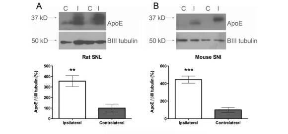
In work we published with Ted Price and colleagues, injury to the sciatic nerve produced a marked increase in APOE. Proteomic analysis showed little detectable APOE in uninjured nerve, followed by strong induction after injury, with evidence of new APOE synthesis within the damaged nerve.[3]
The biological explanation is intuitive:
neural injury creates a sudden demand for lipid recycling and membrane repair, and APOE is part of the machinery mobilized to meet that demand.
This provides an important framework for understanding APOE4 after trauma.
Why APOE4 may reduce resilience after injury
APOE4 differs structurally and functionally from APOE3. These differences affect lipid handling, protein stability, receptor interactions, inflammatory signaling, vascular biology, and communication among neurons and glial cells.
After injury, the nervous system becomes highly dependent on processes in which APOE participates.
A plausible sequence is therefore:
injury → increased demand for lipid transport and membrane repair → altered APOE4-mediated repair → greater persistence of secondary injury
APOE4 does not determine outcome by itself. Rather, it may contribute to why apparently similar injuries have different consequences in different people.
Two other biological pathways strengthen this model: the blood-brain barrier and tau.
APOE4, TBI, and the blood-brain barrier
The blood-brain barrier is the specialized interface separating circulating blood from brain tissue.
Montagne and colleagues demonstrated greater blood-brain-barrier breakdown in cognitively normal APOE4 carriers, particularly in the hippocampus and medial temporal lobe. This was associated with pericyte injury and predicted cognitive decline independently of measured amyloid and tau pathology.[4]
TBI can damage the same neurovascular system. Mechanical forces strain small cerebral vessels and increase blood-brain-barrier permeability, allowing blood proteins and inflammatory signals to enter brain tissue.
The two processes therefore converge:
APOE4-associated vascular vulnerability + trauma-induced vascular injury
This may be one mechanism by which APOE4 alters resilience after head trauma.
Tau provides another connection
Tau normally helps stabilize axons. Mechanical injury stretches axons and can cause tau to detach from microtubules, become abnormally phosphorylated, misfold, and accumulate.
Ojo and colleagues studied repetitive mild TBI in mice expressing human tau. Repeated injury produced persistent axonal damage, gliosis, altered cerebral blood flow, and accumulation of tau and tau oligomers.[5]
APOE4 also appears to influence what happens once pathological tau develops. Experimental models show that APOE4 worsens tau-associated inflammation and neurodegeneration, while removal of neuronal APOE4 reduces tau-related gliosis, neurodegeneration, and propagation of pathology through connected regions.[6]
Human imaging studies point in the same direction. APOE4 carriers appear to enter a phase of accelerated tau accumulation at lower amyloid levels than noncarriers.[7]
Together, these findings support a model in which:
head trauma → axonal damage and abnormal tau → APOE4-associated abnormalities in repair, inflammation, and vascular function → greater persistence or propagation of tau pathology
This pathway has not yet been demonstrated prospectively from concussion to dementia in humans, but several independent lines of evidence converge on the same biology.
What is CTE?
Chronic traumatic encephalopathy, or CTE, is a neurodegenerative disease associated with prolonged exposure to repetitive head impacts. Its defining pathology is abnormal phosphorylated tau deposited around small blood vessels, particularly at the depths of cortical sulci.
CTE is not synonymous with concussion. The strongest association is with long-term cumulative exposure to repetitive head impacts, not with one isolated concussion.[2]
Atherton and colleagues studied 364 brain donors with substantial repetitive-head-impact exposure. APOE4 did not simply determine who had CTE. Among older donors, however, APOE4 was associated with more advanced CTE stage and greater phosphorylated-tau burden. In people aged 65 or older, APOE4 carriage was associated with approximately twice the odds of advanced stage III-IV CTE.[8]
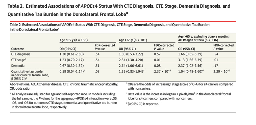
This is consistent with APOE4 acting as a disease modifier: trauma supplies the exposure, while APOE4 may influence the biological response to that exposure.
cPLA2 and the secondary injury response
Another pathway linking neural trauma to neurodegeneration is cytosolic phospholipase A2, or cPLA2.
cPLA2 cleaves membrane phospholipids and releases arachidonic acid. After neural trauma, calcium and inflammatory signaling can activate cPLA2, increasing arachidonic-acid-derived signaling and downstream production of prostaglandins, leukotrienes, and other bioactive lipid mediators.[9]
Arachidonic acid itself is a normal and important component of cell membranes. The concern is not that dietary arachidonic acid is harmful.
Rather, excessive cPLA2 activation after injury may produce an abnormal burst of arachidonic acid signaling that shifts the tissue environment toward persistent or unresolved inflammation.
Experimental studies of TBI and spinal cord injury place cPLA2 within the secondary injury cascade, with effects on membrane integrity, oxidative stress, inflammation, lysosomal biology, autophagy, and neuronal survival.[9]
This has particular relevance to our laboratory because we are developing cPLA2 inhibitors for Alzheimer disease. Whether acute cPLA2 inhibition after TBI can reduce subsequent axonal injury, tau pathology, or neurodegeneration is a testable therapeutic question.
Where omega-3 fatty acids fit
DHA is a major structural fatty acid in neuronal membranes.
Following trauma, membrane lipids undergo rapid remodeling. DHA and EPA also serve as precursors for specialized lipid mediators, including resolvins and protectins, involved in the resolution of inflammation.
This creates an interesting intersection with cPLA2 biology. Excessive cPLA2 activation may increase arachidonic-acid-derived inflammatory signaling, while greater DHA and EPA availability may favor pathways involved in resolution and repair.
Omega-3 fatty acids are not cPLA2 inhibitors, but both approaches influence the lipid environment in which secondary neural injury develops.
Landmark human omega-3 studies
In 2016, Oliver and colleagues randomized 81 NCAA Division I football players to placebo or 2, 4, or 6 g/day of DHA over a 189-day season. They measured neurofilament light, or NfL, a blood marker of axonal injury.
NfL increased during the season, particularly among athletes with greater playing exposure. DHA attenuated that rise when the supplemented groups were considered together.[10]
This provided proof of principle that modifying fatty-acid status might alter a biological marker associated with repetitive head impacts. It did not establish that DHA prevents concussion, tau pathology, cognitive decline, or dementia.
A second randomized study in 2022 tested approximately 2.4 g DHA plus 1.0 g EPA per day in collegiate football players. NfL increased in both groups, and supplementation did not significantly reduce NfL or the inflammatory markers IL-6 and TNF-α.[11]
The individual studies therefore produced heterogeneous results.
A 2024 individual-participant meta-analysis combined three studies involving 179 football players. No significant treatment difference was detected at midseason, but omega-3 supplementation was associated with a smaller postseason increase in NfL.[12]
The overall signal is therefore biologically interesting but not yet clinically definitive.
What about DHA after an actual concussion?
This may be the more relevant therapeutic question.
Miller and colleagues randomized 40 adolescents with sport-related concussion to 2 g/day DHA or placebo, beginning within four days of injury.[13]
The DHA group became symptom-free at a median of approximately 11 days compared with 16 days with placebo and began return-to-sport progression at about 14 versus 19.5 days.
Those differences did not reach conventional statistical significance, but this was a small pilot trial. A roughly five-day numerical separation in recovery provides a rational signal for a properly powered study rather than evidence that the intervention is ineffective.
Why spinal cord injury studies matter
Spinal cord injury provides a useful model because injury timing and treatment can be controlled precisely.
In a landmark experiment, King and colleagues found that omega-3 fatty acids administered shortly after spinal cord injury improved neuronal and oligodendrocyte survival and functional recovery.[14] Subsequent preclinical studies and meta-analyses have generally supported neuroprotective effects of omega-3 treatment, although study quality and publication bias remain limitations.[15]
Some experiments also found that manipulating arachidonic-acid availability altered recovery. This should not be interpreted as evidence that arachidonic acid in the diet is harmful. A more relevant interpretation is that the balance of lipid signaling after injury matters, and excessive cPLA2-driven arachidonic-acid signaling may contribute to unresolved inflammation.
The timing of treatment may be particularly important. In these experiments, omega-3 treatment is not simply improving long-term nutrition. It is being delivered while membrane disruption, inflammation, oxidative stress, and secondary tissue injury are actively evolving.
The missing clinical trial
The major gap is now specific.
A sufficiently powered trial should enroll patients soon after a documented TBI, measure baseline omega-3 status, and randomize them to a defined DHA-rich intervention or placebo.
Clinical recovery should be paired with biological measures such as NfL, GFAP, phosphorylated tau, inflammatory lipid mediators, markers of blood-brain-barrier injury, and advanced MRI.
APOE genotype should be prespecified.
Such a trial could determine whether early DHA modifies secondary axonal injury, whether baseline omega-3 status matters, whether timing affects efficacy, and whether APOE4 carriers respond differently.
The same framework could eventually be used to test cPLA2 inhibition.
What does this mean for APOE4 carriers involved in sports?
A useful distinction is whether head impacts are intrinsic to the sport or occur primarily through accidents.
In American football, rugby, boxing, kickboxing, and mixed martial arts, repeated head impacts are part of normal participation, including many that never cause recognizable concussion symptoms. Cumulative exposure is therefore relevant even when diagnosed concussions are uncommon.
There is no established guideline excluding APOE4 carriers from these sports solely on the basis of genotype. However, reducing repetitive head-impact exposure is biologically consistent with what is known about APOE4, neural repair, vascular vulnerability, and tau pathology.
Cycling, surfing, skiing, snowboarding, and horseback riding are different. Head injury is not intrinsic to participation. It occurs mainly through falls or collisions.
In these activities, individual history becomes especially important.
A cyclist who has ridden for 20 years without a significant crash has a very different exposure history from someone who has fallen four times in a decade and sustained a TBI with loss of consciousness.
The relevant question is therefore not simply whether an activity is classified as a contact sport, but how much meaningful head-impact exposure that individual is actually accumulating.
What can be done after a TBI?
The initial mechanical injury cannot be reversed, but the biological response continues for hours to days.
During this period, the brain undergoes changes in blood flow, energy metabolism, membrane integrity, mitochondrial function, inflammation, oxidative stress, and axonal stability. This secondary-injury window provides a biologically plausible opportunity for intervention.
Current management emphasizes appropriate medical evaluation, avoidance of another injury during recovery, and gradual return to physical and cognitive activity rather than prolonged complete rest.[16]
Long-term risk reduction should also address vascular and metabolic health, including blood pressure, diabetes, smoking, sleep apnea, excessive alcohol use, and physical inactivity.
A Mediterranean-style dietary pattern is reasonable for overall vascular and brain health. Omega-3 fatty acids deserve particular study because they combine a plausible mechanism, substantial preclinical evidence, a human biomarker signal, and an encouraging but underpowered post-concussion trial.
What remains to be established is the optimal dose, timing, duration, baseline omega-3 status, and whether APOE genotype modifies treatment response.
Take-home messages
TBI is a disruption of brain function after trauma, not simply any bump to the head.
APOE participates in neural repair, and APOE4 may reduce resilience through effects on lipid handling, vascular integrity, inflammation, and tau biology.
Cumulative head-impact exposure matters, particularly in football, rugby, boxing, and other sports where impacts are intrinsic to participation.
cPLA2 activation and altered lipid signaling may contribute to secondary injury, while omega-3 fatty acids are a plausible intervention that deserves better acute-TBI trials.
After TBI, the priorities are appropriate recovery, prevention of another injury, and aggressive management of broader vascular and metabolic risk.
References
Centers for Disease Control and Prevention. About Mild TBI and Concussion. CDC.
Centers for Disease Control and Prevention. About Repeated Head Impacts. CDC.
Melemedjian OK, Yassine HN, Shy A, Price TJ. Proteomic and functional annotation analysis of injured peripheral nerves reveals ApoE as a protein upregulated by injury that is modulated by metformin treatment. Mol Pain. 2013;9:14. doi:10.1186/1744-8069-9-14.
Montagne A, Nation DA, Sagare AP, et al. APOE4 leads to blood-brain barrier dysfunction predicting cognitive decline. Nature. 2020;581:71-76. doi:10.1038/s41586-020-2247-3.
Ojo JO, Mouzon B, Algamal M, et al. Chronic repetitive mild traumatic brain injury results in reduced cerebral blood flow, axonal injury, gliosis, and increased T-tau and tau oligomers. J Neuropathol Exp Neurol. 2016;75:636-655. doi:10.1093/jnen/nlw035.
Knoferle J, Yoon SY, Walker D, et al. Neuronal APOE4 removal protects against tau-mediated gliosis, neurodegeneration and myelin deficits. Nat Aging. 2023;3:275-296.
Steward A, Biel D, Dewenter A, et al. ApoE4 and connectivity-mediated spreading of tau pathology at lower amyloid levels. JAMA Neurol. 2023;80:1295-1306.
Atherton K, Han X, Chung J, et al. Association of APOE genotypes and chronic traumatic encephalopathy. JAMA Neurol. 2022;79:787-796. doi:10.1001/jamaneurol.2022.1634.
Zhang HJ, Chen YT, Hu XL, et al. Functions and mechanisms of cytosolic phospholipase A2 in central nervous system trauma. Neural Regen Res. 2023;18:258-266. doi:10.4103/1673-5374.346460.
Oliver JM, Jones MT, Kirk KM, et al. Effect of docosahexaenoic acid on a biomarker of head trauma in American football. Med Sci Sports Exerc. 2016;48:974-982. doi:10.1249/MSS.0000000000000875.
Mullins VA, et al. Effects of fish oil on biomarkers of axonal injury and inflammation in American football players: a placebo-controlled randomized controlled trial. Nutrients. 2022;14:2139.
Heileson JL, Macartney MJ, Watson NL, et al. Nutritional optimization for brain health in contact sports: a systematic review and meta-analysis on long-chain ω-3 fatty acids and neurofilament light. Curr Dev Nutr. 2024;8:104454.
Miller SM, Zynda AJ, Sabatino MJ, Jo C, Ellis HB, Dimeff RJ. A pilot randomized controlled trial of docosahexaenoic acid for the treatment of sport-related concussion in adolescents. Clin Pediatr (Phila). 2022;61:785-794.
King VR, Huang WL, Dyall SC, Curran OE, Priestley JV, Michael-Titus AT. Omega-3 fatty acids improve recovery, whereas omega-6 fatty acids worsen outcome, after spinal cord injury in the adult rat. J Neurosci. 2006;26:4672-4680.
MacIntosh-Smith WAC, Abdallah A, Cunningham CJ. The potential effects of polyunsaturated ω-3 fatty acids on spinal cord injury: a systematic review and meta-analysis of preclinical evidence. Prostaglandins Leukot Essent Fatty Acids. 2023;191:102554.
Patricios JS, Schneider KJ, Dvorak J, et al. Consensus statement on concussion in sport: the 6th International Conference on Concussion in Sport, Amsterdam 2022. Br J Sports Med. 2023;57:695-711.
Disclosure: This post contains partially AI-assisted text. All references were reviewed for accuracy. I have no relevant conflicts of interest. These opinions are mine and do not reflect the opinions of my employer.
Your point that a normal CT doesn't rule out concussion is the one is one every APOE4 carrier to hear. In the ER, the head CT is looking for bleeding that needs a surgeon. It's always a tough conversation when parents push for head CT's on kids that only had minor bumps as we have to explain that, short of a bleed, CT scans don't show concussions. And then the risk benefit conversation has to occur re: radiation exposure that may be unnecessary.
But, it cuts both ways, when it comes back clean, a lot of people go home thinking they've been cleared, when the CT can't look for concussion at all. So, if you carry APOE4 and you've hit your head, your symptoms are the diagnosis.
Either way, CT or no CT, get the return-to-activity guidance in writing and take the recovery time seriously, especially before getting back on the bike.